An important update is available for FreeStyle LibreLinkØ. Check here for more information.
27 Sep 2018

There are many types of controls required to execute a flow cytometric experiment effectively. These cover all areas from controls to determine validity of reagent staining, through to controls that are required to effectively analyse data. However one must not forget that the key part of any experiment, no matter how well controlled, is sample quality. Many factors can affect the quality of your sample and therefore have an impact on expected results. These include:
Flow cytometry buffers can vary from lab to lab, but mostly commonly, the standard staining buffer is PBS with serum added (FCS (1-10%) or BSA (0.1-1%)). Serum proteins protect cells from apoptosis, prevent non-specific staining and also prevent cells from sticking to the side of your tubes. Depending on your cell type it may also be advisable to add EDTA to prevent cation based cell to cell adhesion as flow cytometry aims to assess single cells (important for digested tissue samples). Clumps can also block your cytometer! However, be sure to first titrate the concentrations of EDTA in case your cells are sensitive to it. Also check that none of your antibodies are dependent on the presence of cations!
Single cell suspensions must be prepared for cytometric analyses, and some starting material is easier to manage than others. Blood, for example is all ready to go, however obtaining single cell suspensions from solid tissues can be much more complex. Key things to remember when digesting tissues:
Temperature and staining duration can also have an effect on the quality of your data. For example, staining at room temperature may reduce the antibody dose and staining duration, but viability may drop and non-specific staining may increase. Therefore make sure all reagents are titrated and tested under the same conditions as your final experiment.
On an ending note, the key to flow cytometric success is to make sure all bases are covered, from starting with high quality samples, to using the correct controls and optimising conditions and reagents.
Dr Anna Brooks holds a BCA (management) and a PhD in Immunology and is a Senior Research Fellow with the Maurice Wilkins Centre at the University of Auckland. Anna is also an expert flow cytometrist and is director of Auckland Cytometry, the flow cytometry core facility for the Faculty of Science. Anna’s primary interest lies in developing multicolour panels to characterise complex cellular populations in digested human tissues. Anna is also an active member of the international flow cytometry community and currently sits on the Australasian Cytometry Society Council as research liaison. When not marvelling in the many fluorescent colours of the cytometry world, Anna enjoys to scuba dive and explore the vibrant wonders of our underwater world.
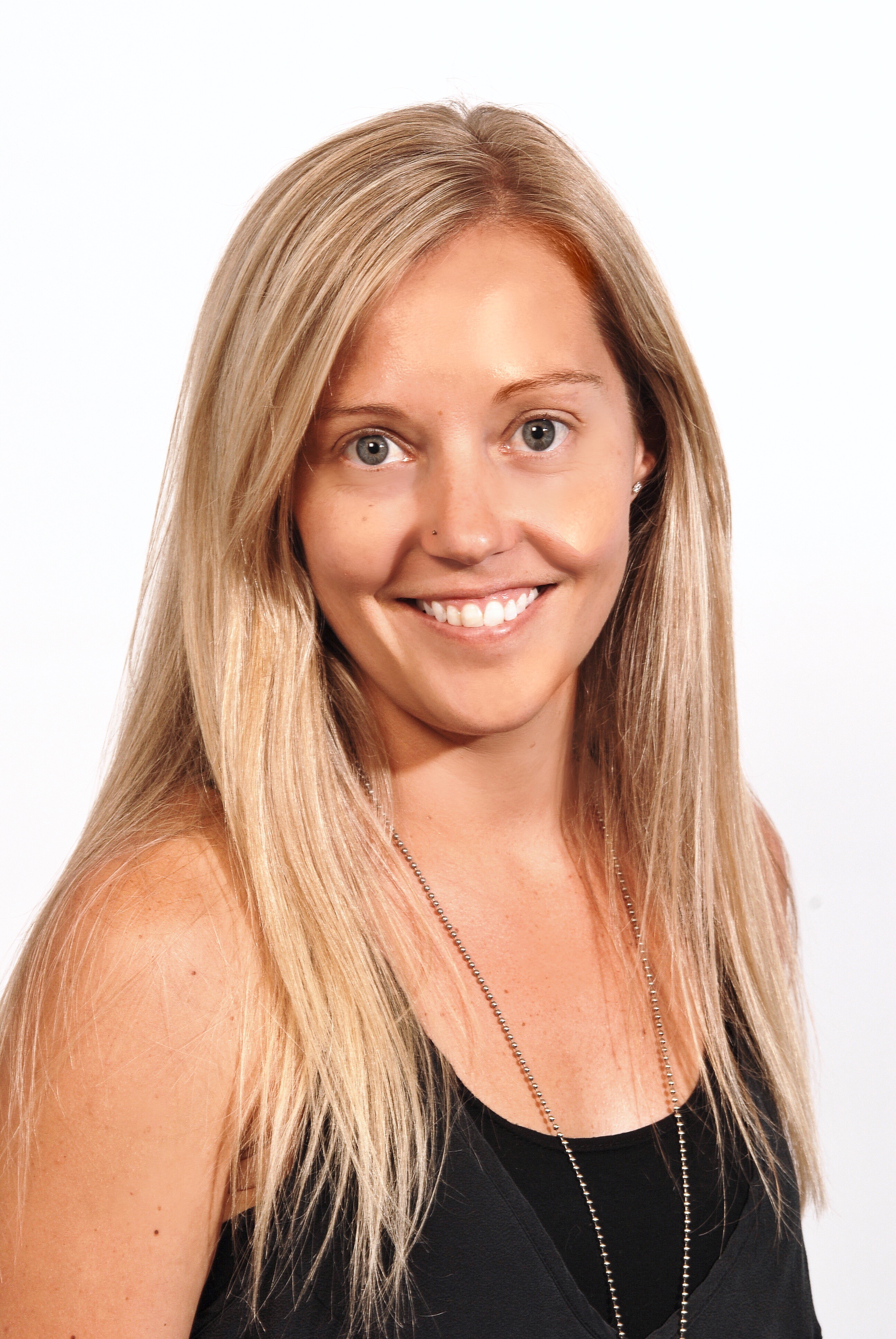
Dr Anna Brooks
University Of Auckland, School Of Biological Sciences
27 Sep 2018
If you enjoyed reading our articles, why not sign up to our blog mailing list? You'll get new articles straight to your inbox as they're released!