The µMACS and MultiMACS Protein A/G Kits contain colloidal super-paramagnetic MicroBeads, which are conjugated to Protein A/G, respectively. The non-sedimenting Protein A/G MicroBeads ensure rapid reaction kinetics enabling the formation of the labeled immune complex in only 30 minutes. Compared to standard protocols, on-column IPs result in reduced non-specific background binding and less protein dissociation from the complex. Moreover, laborious centrifugation or buffer removal steps can be avoided.
Detailed procedure
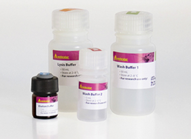
After cell lysis the lysate is incubated with the specific antibody or serum against the target protein. Pre-clearing of the lysate is dispensable. Then the antibody-protein complex is magnetically labeled with µMACS Protein A/G MicroBeads. The sample is loaded onto a MACS Column placed in the magnetic field of the µMACS or thermoMACS Separator. After washing, the magnetically labeled antibody-protein complex and associated molecules are retained on the column. After elution the (co-)immunoprecipitated proteins can be analyzed by SDS-PAGE and Western blot.
Applications
The advantages of µMACS Protein A/G MicroBeads for ChIP are shown in a customer protocol and have been incorporated into a special protocol. In contrast to the standard ChIP protocol, this streamlined protocol saves 90% of laboratory time in particular on pre-clearing, incubation times, and reverse crosslinking steps. In addition, it allows for working with limited starting materials of just 106 cells. Moreover, customer data indicated enhanced ChIP and a significantly reduced level of non-specific PCR products.
Downstream applications
The (co-)immunoprecipitated proteins can be analyzed by SDS-polyacrylamide gel electrophoresis and subsequent Western blotting techniques or by on-column enzymatic reactions using the thermoMACS Separator.